INTRODUCTION

Computer vision is a branch of Artificial Intelligence / Machine Learning (AI/ML) that tries to mimic the tasks performed by human vision. For computers, it means processing digital image and video (sequence of images) to detect objects or patterns, identification of objects, and their classification etc.

Work done by a Radiologist often includes similar tasks i.e., finding a pattern of irregularity in bones or tissues. This makes Computer Vision ideal choice for helping radiologist by automating some of the tasks, thus improving productivity.

Benefits of using AI Computer Vision technology

N

Increase in productivity of Medical Practitioners

N

Screening and Triaging of images waiting in queue

N

Automation of clinical records

N

Reduce mistakes in manual checking of image

N

Monitoring and quantification of diseased area across timeframe

EVOLUTION OF COMPUTER VISION

Although Computer vision as a technology existed from 1970s, the initial techniques were largely based on image processing and mathematical models that helped perform basic tasks like detecting edges in the image, separating the object from the background and segmentation. Real boost to computer vision came with advent of deep learning frameworks from 2010. Deep Learning based algorithms helped in finding more complex patterns without explicitly defining the pattern. Since then, there have been rapid advances in deep learning based Computer Vision. The problem such as classification of Dog and Cat images, that seemed challenging just a few years back has now become ‘Hello world’ exercise of today.

With Deep Learning based computer vision models routinely scoring more than 90% accuracy, tech community quickly adapted Computer Vision in every domain that requires image or video analysis. Some of the obvious use cases are face detection, security surveillance, Self-driving car, Defense, OCR, gaming, and Healthcare.

MEDICAL IMAGING

Medical imaging is a non-invasive technique of imaging interiors of patient’s body for clinical analysis. This technique is often used to examine bones and tissues for any abnormality. Interpretation of the images is done by a qualified Radiologist.

Most common imaging techniques are

N
X-Ray.
N
Computed Tomography (CT).
N
Magnetic Resonance Imaging (MRI).
N
Ultrasound.

DIGITAL IMAGING STANDARDS IN MEDICAL IMAGING

N
DICOM (Digital imaging and Communication in Medicine) is used for storing and exchanging medical images. Typical DICOM file contains a header and image. Header stores metadata of patient and imaging technique used.
N
PACS (Picture Archiving and Communication System) is a system used for storing and exchanging image files. PACS is used for implementing workflow of images from capture to analysis to archiving.

TYPES OF COMPUTER VISION ALGORITHMS USED IN MEDICAL IMAGING

N
Classification: The classification algorithm will classify the image in two or more classes. For example, algorithm trained to detect lung nodule can classify chest X-ray based on presence of absence of a nodule
N
Localization: In Localization, the algorithm will point to location of the pattern by drawing bounding box around the problematic area. For example, in mammography, algorithm can draw box around suspected tumor on breast X-ray image
N
Segmentation: It is a technique that can color code set of pixels in image representing area of the pattern detected. This is often used to determine size of the abnormality. Comparing different images taken at different times can show the change in size of abnormality.

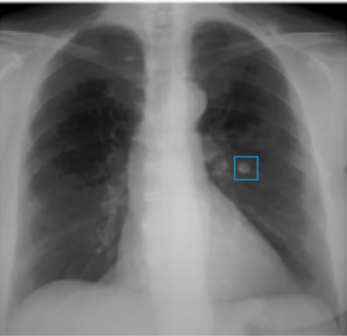
Figure 1: Chest X-ray showing localization of abnormality.2

Role of AI in Medical Imaging

Computer Vision model that is trained on required medical images can be used to identify, classify and locate a specific pattern e.g. Fractured bone or a tumor. Although computer vision algorithm can show high accuracy in analyzing the medical image, it is rarely used to replace a role of actual radiologist or pathologist.

Typical use cases of AI in medical imaging includes

N
Diagnostic Assistance: Radiologists often examine hundreds of images per day. AI algorithm can assist Radiologist by saving time spent in diagnosis by highlighting the suspicious area of the image or by quantification of the area.
N
Screening and Triaging: When there is a long queue of images to be checked by Radiologist, AI algorithm can analyze and triage the images in PACS, so that most critical cases get early attention.
N
Monitoring: To analyze response to the treatment (e.g. in Oncology), images of diseased tissues captured at different times are aligned and compared. The change in size of diseased tissue can help in understanding the response to the treatment.
N
Charting: Post review of findings made by AI tool by the medical practitioner, typical AI product often provides the necessary inputs required for clinical charting that otherwise need to be manually recorded.

Typical application of Computer vision in Imaging

N
Cardio-vascular image analysis .
N
Oncology: Tumor detection and monitoring.
N
Ophthalmology: Detection of diabetic retinopathy.
N
Neurology: Stroke Detection in CT.
N
Orthopedic: Detection of fractured bone.
N
Emergency medicine: Triage and diagnosis of time sensitive patients.
N
MRI brain interpretation.
N
Chest X-ray assessment for Pneumonia detection.
N
Dental: X-ray Image annotation and diagnosis .

Critical Success Factor

N
Accuracy: AI Products are already trained on extensive data. However, all Deep Learning based products have ‘bias’ based on data used for training. It is important to validate if the AI/ML product is correctly predicting for every new installation.
N
Seamless integration: AI product cannot exist in isolation. It needs to be integrated seamlessly in application ecosystem. The integration should be well designed to save time. You do not want Doctor to stare at screen for long time till images are getting uploaded.
N
Training: When medical practitioners are using the product, they are also training the product. It is important to educate them for correct usage of the product.
N
Productivity Metrics: Business case is normally based on increase in productivity. To validate the increase in productivity, metrics should be built in the integration or within the AI Product.
N
Data Security: AI/Computer Vision technology is well democratized. There are good AI product companies in several countries. Though AI vendor is registered in your country, it is important to check where the product is hosted, where developers who have access to your data are based at.
Computer Vision based AI/ML algorithms can be used in medical imaging to boost the productivity of medical practitioners. To large extent these applications can help in diagnosis. At least today, these AI products cannot replace the Radiologist or Pathologist. There is also a factor of regulatory agencies like FDA allowing these products to be used in diagnosis.
Sources used for this blog-
1. Benjamens S, Dhunnoo P, Meskó B. The state of artificial intelligence-based FDA-approved medical devices and algorithms: an online database. NPJ Digit Med.2020 Sep 11;3:118. doi: 10.1038/s41746-020-00324-0. PMID: 32984550; PMCID: PMC7486909.
2. Udacity: AI for Healthcare

Newscape Consulting has been working with several AI products utilizing Computer Vision. Newscape can be a valuable partner in selecting, piloting and implementing AI products.

Share